Dear Echocardiographers,
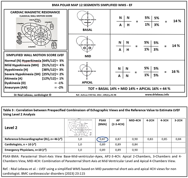
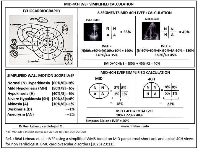
In the era of Point of Care Ultrasound Study (POCUS) , we have recently published an article describing a novel method to predict left ventricular ejection fraction (LVEF). We developed a 4-segments wall motion score (WMS) model using the parasternal short axis view at the papillary muscle (MID) and the apical 4 Chambers (4CH) views. (Cf ref 1 and fig 1 )
The MID and 4 Chambers views are some of the most acquired by non cardiologist ( emergency physician, critical care physician, anesthesiologist…) . The advantage of these two views is that they are both perfused by all 3 coronary arteries . Furthermore the apical 4-chambers view allows for a good analysis of apical abnormalities (dyskinesia and aneurysm)
Real Lebeau md
Cardiologist echocardiographer
Hospital Sacre-Coeur Montreal Canada
MID-4CH LVEF
BMA Polar Map 12 segments Simplified WMS - EF